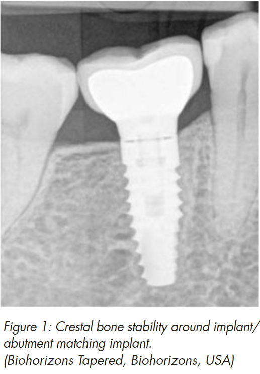
Crestal bone stability around dental implants remains one of the most important and wanted features of successful implant treatment. Besides major clinical advantages to the patient, stable marginal bone provides us, the clinicians, with psychological comfort and satisfaction, because of positive long-term outcome (Fig 1). Therefore we all need to be aware of possible causes, which can lead to loss of crestal bone stability and exercise every method to prevent bone resorption.
For almost one decade platform switching was considered to be the most effective way to achieve this task. It was so effective that almost all implant companies have implemented platform switching as an essential feature of implant manufacturing. Everybody made the conclusion that implant design was more important that the biology itself. However, recent clinical research of our group has shown that soft tissue thickness is an important factor in preserving crestal bone stability around implants. It was determined that if vertical soft tissue thickness is 2 mm or less, there will be crestal bone resorption of 1.5 mm extent during formation of biological seal between soft tissues and implant/abutment/restoration surfaces (Fig 2).
Furthermore, it was clearly shown that even implants with platform switching modification couldn’t maintain bone, if at the time of implant placement, vertical soft tissues were thin (Fig 3). That brings the discussion – what is more important, biology or implant design back on the table. Well, to continue further, we need to understand, that vertical soft tissue thickness is the prerequisite of the biological width around implants. Biological width around implants starts to form at the time of healing abutment connection and is completely finished after 8 weeks. This biological seal is the only and most important protection barrier of the osseonintegrated implant from contaminated intraoral environment. Thus there is a direct connection between pre-implant mucosa of edentulous alveolar ridge and peri-implant soft tissues.
It seems that soft tissue thickness required to protect underlying bone around implants is approximately 4 mm, which is wider than the biological width around teeth. There are 2 ways how biological width around implants can be formed: with crestal bone loss or without bone resorption. Which one would you like to your patient to have? Or which one would you like your mother to have? That is the question we all as clinicians should answer sincerely.
So if we diagnose thin vertical tissues at the time of implant placement, what should we do? There are no guidelines to follow so far, however we need to do something, because crestal bone resorption will otherwise result. This is especially important for short implants, which usage is becoming common practice. Today, implants in 8mm length are no longer considered short anymore, and we have sufficient data to say, that 6 mm length implants work as good as longer ones in posterior areas of both jaws. However, imagine the outcome if a 6mm implant is placed in mandibular posterior jaw region, where thin vertical soft tissues are frequently present. We would have approximately 2 mm of bone resorption, due to biological width formation. It leaves only 2/3 of implant surface osseonintegrated. Now, that is a risk of implant failure, keeping in mind the prosthetic suprastructure and implant/crown ratio. Some implant manufacturers have launched 4 mm length implants, therefore soft tissue thickness is even more important for the users of these products.
So what is the strategy? There are several options, some of them already researched clinically; some of them are based on clinical expertise without any serious evidence. The initial thought is to just place the implant deeper sub-crestally (Fig 4). Firstly, there must be enough distance to the alveolar nerve that the implant can be positioned sub-crestally in a safe manner. It is advised that the implant would stop at least 1 mm over the nerve.


Extensive sub-crestal positioning of the implant, of course, does not prevent crestal bone loss, as the microgap at the implant-abutment interface will form an inflammatory infiltrate, which will resorb the bone anyway; however it is likely that the implant will not have soft tissue recession or rough surface exposure, which usually follow bone resorption. It is well known that the exposure of the rough implant surface enhances plaque accumulation and development of peri-implantitis.
Consequently, we might use the third option – vertical reconstruction of soft tissue thickness, which in my opinion is the most logical approach. Increasing soft tissue thickness vertically compensates the lack of vertical tissue thickness. Already in a JOMI 2009 paper “The influence of soft tissue thickness on crestal bone changes around implants: a 1-year prospective controlled clinical trial” we have suggested that clinicians “… consider the thickening of thin mucosa before implant placement”, therefore this concept is not entirely new. The idea is to place some sort of autogenic, allogenic or xenogeneic material over the implant in that way increasing soft tissue thickness after healing.
A connective tissue graft is considered the golden standard for soft tissue augmentation around implants. However this technique has some serious disadvantages, like donor site morbidity and difficulty of the harvesting procedure. Therefore allogenic substitutes might be considered as a viable option to replace autogenous grafts in vertical soft tissue reconstruction. The use of accelular dermal matrix so far is the only approach backed by solid clinical research, including a controlled clinical prospective study.
In this study, implants were placed in 3 groups of patients with (1) thin vertical tissues, (2) thick vertical tissues and (3) thin vertical tissues augmented with acellular dermal matrix material. Radiographic assessment showed the reduction of crestal bone loss from 1.74 mm in the thin tissue group to 0.32 mm in the augmented group. In addition, the increase of soft tissue thickness went up 2.33 mm – from 1.50 mm to 3.83 mm after augmentation with allograft (Fig 5ab). This research proves that the lack of vertical soft tissue thickness required for biological width formation without crestal bone loss can be compensated by the use of accelular dermal matrix material at the time of implant placement.
In conclusion it must beemphasized that diagnosis of thin vertical soft tissues is very important point in implant treatment. Only by acknowledging the fact that tissue thickness is an important factor, we can use the protocols, which allow us to reconstruct vertical peri-implant tissues and reduce crestal bone loss.
Dr Tomas Linkevicius, DDS, Dip Pros, PhD Private Practice, Vilnius, Lithuania. Contact: linktomo@gmail.com
References
1. Linkevicius T, Apse P, Grybauskas S, Puisys A. The influence of soft tissue thickness on crestal bone changes around implants: a 1-year prospective controlled clinical trial. Int J Oral Maxillofac Implants. 2009 Jul-Aug;24(4):712-9. 2. Puisys A, Vindasiute E, Linkeviciene L, Linkevicius T. The use of acellular dermal matrix membrane for vertical soft tissue augmentation during submerged implant placement: a case series. Clin Oral Implants Res. 2015 26(4) 465-470
Reprinted with permission from International Dentistry - Australasian Edition
Linkevicius T. Int Dent Aus Ed 2018;3:14-16
Dr Linkevicius will be presenting at the 10th International Quintessence Symposium on Periodontics & Restorative Dentistry | New Frontiers of Aesthetic Excellence | Successfully Integrating the Best of Traditional & Digital Dentistry. To register visit www.iqs.sydney